Hybrid MRI image‑enhancement approaches that combine traditional reconstruction methods with deep learning can improve image quality and efficiency, but radiographers play a critical role in selecting, evaluating, and safely implementing these tools in daily practice.
Introduction
Magnetic resonance imaging (MRI) image quality has a direct impact on diagnostic confidence, workflow efficiency, and patient experience. Radiographers routinely manage challenges such as noise, motion artefacts, and limited spatial resolution. While no single enhancement method performs optimally in all situations, combining traditional MRI reconstruction techniques with deep learning approaches offers clinically relevant improvements.
This blog summarises key insights from a recent scoping review (available in Journal of Clinical Radiography and Radiotherapy Vol. 24) on MRI image enhancement and translates them into practical, practice‑oriented knowledge for radiographers and imaging professionals.
Why MRI Image Enhancement Matters in Practice
MRI is valued for its excellent soft‑tissue contrast, but image quality can be compromised by motion, long acquisition times, and system‑dependent limitations. These challenges affect diagnostic accuracy, examination efficiency, and patient tolerance. Current MRI research increasingly focuses on reducing artefacts and noise while preserving diagnostically meaningful anatomy within workflows that are feasible in routine clinical practice.
Traditional Enhancement Techniques: Still Relevant
Conventional MRI image‑enhancement methods remain fundamental in many clinical settings. Techniques such as non‑local means (NLM) filtering, motion correction strategies, parallel imaging, and optimised k‑space sampling have demonstrated effectiveness in reducing noise and artefacts. These approaches are grounded in MRI physics and provide transparency and predictable behaviour, although they may show limitations in highly accelerated or low‑signal scenarios.
The Role of Deep Learning in MRI Enhancement
Deep learning‑based reconstruction and enhancement methods have shown strong performance in denoising, super‑resolution, and motion compensation. Convolutional neural networks can learn complex artefact and noise patterns, enabling faster acquisitions and improved signal‑to‑noise ratio, particularly in diffusion‑weighted and accelerated imaging protocols. Many of these solutions are now integrated into commercial MRI systems.
At the same time, deep learning methods raise important considerations related to generalisability, validation, and interpretability. Over‑smoothing or alteration of image texture remains a concern, highlighting the need for careful clinical evaluation.
Why Hybrid Approaches Show the Most Promise
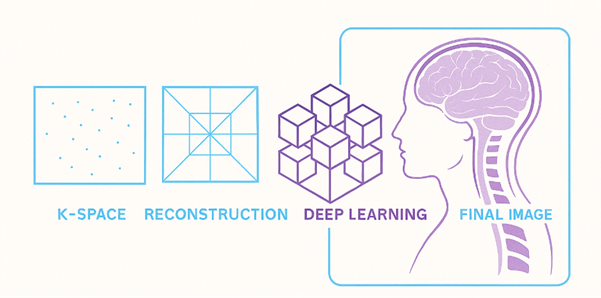
A central conclusion of contemporary research is that hybrid approaches—combining traditional MRI reconstruction techniques with deep learning—offer the most balanced and clinically robust solution. Traditional methods provide physics‑based constraints, while deep learning contributes efficiency and enhanced performance. Together, they reduce reliance on black‑box algorithms while delivering meaningful improvements in image quality.
Practical Takeaways for Radiographers
For radiographers, these findings translate into practical, learnable actions that can be applied directly at the scanner and during protocol development:
- Protocol optimisation: During protocol setup, compare deep learning reconstructions side‑by‑side with conventional reconstructions. Assess signal‑to‑noise ratio, edge sharpness, contrast behaviour, and preservation of fine anatomical detail.
- Efficient scan strategies: Use AI‑supported or accelerated reconstruction options to shorten acquisition times when patient comfort, motion risk, or workflow efficiency is a priority, while ensuring diagnostic adequacy.
- Image quality assessment: Routinely evaluate images for potential effects of enhancement, such as over‑smoothing, altered texture, or loss of subtle structures, especially in diffusion‑weighted or highly accelerated sequences.
- Motion‑robust imaging decisions: Continue to apply traditional motion‑reduction strategies (patient positioning, coaching, padding, gating) in combination with advanced reconstruction rather than relying solely on post‑processing enhancement.
- Professional communication: Clearly communicate to radiologists which reconstruction or enhancement method was used and how it may influence image appearance, supporting confident interpretation and interdisciplinary collaboration.
These practices reinforce the radiographer’s role as an informed decision‑maker who actively shapes image quality and patient‑centred MRI care.
Limitations and Future Directions
Much of the advanced MRI enhancement research originates from high‑resource settings, which may limit generalisability. Future work should prioritise validation across different scanners, protocols, and patient populations, as well as improving transparency in deep learning models. Continued collaboration between researchers, radiographers, clinicians, and vendors is essential to translate innovation into reliable clinical benefit.
Conclusion
MRI image enhancement continues to evolve through advances in both traditional reconstruction techniques and deep learning methods. While no single approach is universally optimal, hybrid strategies offer a practical, evidence‑based path forward. For radiographers, engagement with this research supports professional development and contributes to the delivery of safe, high‑quality, and patient‑centred MRI services.
Author Muyiwa (Muyi) Olabode, PhD is a research and development engineer and researcher with expertise spanning MRI‑related imaging research, medical devices, and health‑technology innovation. He works across industry and academia to translate advanced engineering and imaging research into practical, evidence‑based solutions that support radiographers, clinicians, and patient‑centred care.
References
Olabode, M., Tan, L. X. R., Tan, N. K. Y., Mohamed Fauzan, I. F., Törnroos, S., & Lian, C. (2026). MRI Image Enhancement: A Scoping Review of Traditional Techniques and Deep Learning Approaches. Journal of Clinical Radiography and Radiotherapy, 24.
McRobbie, D. W., Moore, E. A., Graves, M. J., & Prince, M. R. (2017). MRI from Picture to Proton (3rd ed.). Cambridge University Press.
Manjón, J. V., Coupé, P., Martí‑Bonmatí, L., Collins, D. L., & Robles, M. (2010). Adaptive non‑local means denoising of MR images with spatially varying noise levels. Journal of Magnetic Resonance Imaging, 31(1), 192–203.
Knoll, F., Hammernik, K., Zhang, C., et al. (2020). Deep‑learning methods for parallel magnetic resonance imaging reconstruction: A survey. IEEE Signal Processing Magazine, 37(1), 128–140.
Aggarwal, H. K., Mani, M. P., & Jacob, M. (2018). MoDL: Model‑based deep learning architecture for inverse problems. IEEE Transactions on Medical Imaging, 38(2), 394–405.